 |
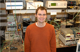
| From Postdeadline paper CTh5D.1 "Wavelength-Size Silicon Modulator." Scanning electron micrograph of the silicon integrated waveguide modulator. |
Make sure to stretch your legs if you want to move from session to session in this frenzy of fantastic photonics research (say that five times fast). Tonight from 8:00-10:00 pm marks the crème de la crème of contributed papers to CLEO. I haven't quite made up my mind of which to attend, but found a number of them particularly exciting:
CTh5D.1, "Wavelength-size Silicon Modulator" from V.J. Sorger et al.
This is work out of the Zhang Lab from Berkely showing an optical modulator with 1-dB/micron extinction (a 20 micron long device gives 20 dB extinction). The modulator is based upon tuning the carrier concentration of an active nm-thin layer of Indium Tin Oxide sandwiched between a MOS structure. Just yesterday, Larry Coldren from UCSB was gently ribbing the silicon folk in his tutorial CW1k.1, "Single-chip Integrated Transmitters and Receivers" for the dearth of practical active components such as a modulator. Coldren sees InP based photonic circuits as the more robust platform for photonic integrated circuits. However, great work like this from the Zhang group will be pushing silicon to the forefront.
CTh5C.4, "In Vivo Three-Photon Microscopy of Subcoritical Structures wihtin an Intact Mouse Brain" from N. Horton et al.
This work from the Xu Group from Cornell University uses a clever choice of longer-excitation wavelength coupled to the improved localization of three photon fluorescence in order to image deep through intact tissue. Even though longer wavelengths are more readily absorbed in tissue, they are significantly less scattered. The overall effect is higher throughput and deeper penetration. Combine that with a 1/z4 fall-off in three-photon fluorescence signal (tighter localization), and now you can make beautiful images of intact tissue. The Xu group shows 1.2 mm stack of brain tissue taken in 4 micron increments. The broader goal will be to eventually use this for optical biopsy in humans. I would prefer to have my tissue scanned with a laser rather than excised from my body with a knife by a surgeon, wouldn't you?
CTh5C.1, "Demonstration of a Bright 50 Hz Repetition Rate Table-Top Soft X-Ray Laser Driven by a Diode-Pumped Laser" from B. Reagan et al.
This work from the Rocca Group of Colorado State University and the Research Center for Extreme Ultraviolet Science and Technology shows a significant improvement of table-top soft x-ray lasers. To see how quickly this group is improving these systems, just look at a March 2012 Laser Focus World feature article highlighting their work- now outdated. The aim of table-top soft x-ray research is to bring systems that are typically found at a shared national lab facility to the many optics tables of university labs and industry. Applications for coherent soft x-rays include laser-induced materials processing at the nanoscale level as well as ultrafast characterization of nanoscale motion. Spectra Physics or Coherent may not be selling ultrfast soft x-ray lasers just yet, but this paper shows a 5-fold increase in repetition rate (important for higher average power applications) and a 20-fold increase in pulse energy from previous best efforts.
ATh5A.4 "Highly Efficient GaAs Solar Cells with Dual Layer of Quantum Dots and a Flexible PDMS Film" from C. Lin et al.
In this paper a Taiwanese collobaration from the Institute of Photonic Systems, National Chiao Tung University, and the Research Center for Applied Sciences has shown a 22 % enhanced efficiency in a GaAs solar cell by spraying a coating of UV absorptive quantum dots onto a polydimethylsiloxane film at the top surface of the cell. This collaboration has found a clever way to not let so many UV photons from the sun go to waste.
No comments:
Post a Comment