
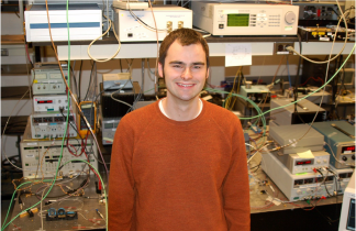
Image from Webb Lab, Cornell University, Adapted by J. van Howe. Left: One photon fluorescence in a fluoroscein solution. Right: two-photon fluorescence in the same solution.
At the very first conference I attended as a new graduate student, I asked my advisor, "So what talks are you going to?" To my surprise he said, "Talks? I don't go to any talks, I catch up with my friends and colleagues and go to the expo to get ideas." My graduate advisor was exaggerating. He did, and still does, go to talks, but his advice then, which I still take to heart, made me realize how important it is to talk to colleagues after and in between talks, as well as the importance of the expo. From an academic point of view, the expo typically is a place to buy new tools to further on-going research. From from my graduate advisor's point-of-view it is also a place to find strengths and weakness in developing technology, the latter typically being more helpful for beginning new research.
As I was scouting companies to find out more information on Mid-IR laser sources (by the way IPG photonics and Daylight Solutions seem to be leading the market in sources in this spectral range), I learned about the impact multiphoton microscopy (MPM) has had on the sales of Ti:Sapph and OPO systems from Stephen Knapp of Coherent, Inc. In fact, there will be Market Focus talk, in the Biophotonics session on Thursday on the showroom floor from Arnd Krueger of Spectra Physics regarding this very subject. Also, be sure to check out an entire session devoted to MPM work, today at 1:30 pm in room A4, or check out the abstracts on the conference CD if you miss it.
A Ti:Sapph plus an OPO is not cheap. You're talking around $200k or more depending on options. These bulk solid state systems used to be the purview of only laser jocks. However, companies like Coherent and Spectra Physics are making them more into turn-key systems in order to put them into the hands (or onto the optical tables anyway) of biologists and biomedical researchers. Though companies like Coherent and Spectra Physics are leading the way of making these sources turn-key, the fact that they are still very expensive and fairly large makes us fiber laser specialists excited. We think the next generation of MPM sources will be all fiber-based and therefore truly compact, turn-key, and a fraction of the price.
To my surprise, Stephen told me that Coherent will sell up to 50 a quarter, mainly for use in MPM! So what is so great about MPM? Well, it is arguably the leading technique for deep-tissue imaging. Being in Chris Xu's group at Cornell University when I was a graduate student, who helped pioneer two-photon microscopy with Winfred Denk, and Watt Webb when he himself was a Cornell grad, MPM buzz has rubbed off on me and I can't miss an opportunity to say something about it. Particularly, I wanted to share one of my favorite photos taken in the Webb lab (above) that gets right to the essence of how MPM works.
The goal of MPM is to get rid of the scattering background outside of the focal plane, the photo on the left. These photons are noise and do not contribute to meaningful information about the sample. Using two-photon fluorescence, however, one gets photons mainly near the focal plane and nowhere else, thereby significantly reducing the background. This allows the biomedical researcher, or surgeon, to scan deep (~1 mm) through tissue. Reduction in scattering occurs because in the two-photon configuration, the probability of a fluorescence event is much greater at the focus of the objective than anywhere else.
The two-photon cross section of typical fluorophores is around 30 orders of magnitude smaller than the one-photon cross section. In order to get a two-photon event, photons must arrive "simultaneously." "Simultaneously" can be figured out from the Heisenberg uncertainty principle to be about 0.5 fs - just use the energy of the transition and solve for time. In order to get a two-photon event, one needs to bunch photons together to stack probability. The microscope objective does this in space and a pulsed excitation source does it in time. The focus is the most probable spot for excitation since photons are bunched in both space and time at this point and no where else. This is why you need a pulsed excitation source for MPM. For more details on MPM see the Dr. Bio webpage at Cornell.

Image from Webb lab and Nikitin lab at Cornell University. Left: H&E stained histology of sliced ovary. Right:Multiphoton image of a follicle within an unstained, intact ovary from mouse. Autofluorescence (green) derives from NAD(P)H and retinol within the tissue. SHG (red) delineates the bursa.
Above are some more pretty pictures of nonlinear microscopy in action. Note that on the left is a typical histology of a mouse ovary. The tissue has been excised, and then stained. On the right shows two-photon microscopy combined with second harmonic generation microscopy of an intact mouse ovary. The moral of the story is the one on the right required no cutting and therefore is much less invasive.
Great post!
ReplyDeleteI've been doing a bunch of research in MPM, evaluating the potential of using adaptive optics to improve the images. The Lin lab at Mass. General Hospital has done some imaging of mice and Jennifer Hunter, up at University of Rochester in David Williams' lab showed really impressive images this year at ARVO.
One question (to you, or any other readers): Do you know of any other types of lasers being used for this? I know all I am aware of are the Ti:Sapphs you mentioned.
Thanks for exposing this exciting discipline and opening my eyes to some more of technical detail and technology surrounding it!